Atelectasis
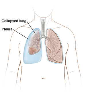
Atelectasis is the collapse of one or more sections (lobes) of the lungs. When you breathe in, the lungs normally expand to fill with air. With atelectasis, a blockage or pressure in the area around the lung (pleura) keeps the lung from expanding. This condition can also occur when the lungs don't have enough surfactant to expand. Surfactant is a substance that forms a thin layer in the lungs to help keep the air sacs open. If your lungs don't fill with air, they can't deliver enough oxygen to your blood.
What causes atelectasis?
Atelectasis is caused by a blocked airway, or pressure from outside the lung. Some causes include:
-
An object or tumor in an airway
-
Mucus blocking an airway
-
Certain lung diseases, including infections
-
Fluid or air buildup in the space around the lung (pleura). Fluid buildup in the pleural space is called a pleural effusion. Air buildup in the pleural space is called a pneumothorax.
-
General anesthesia for chest and belly (abdominal) surgery combined with opioid medicines for pain control
You may also be at higher risk for atelectasis if you are obese or have sleep apnea, asthma, COPD, cystic fibrosis, or other lung disease.
What are the symptoms of atelectasis?
If only a small part of your lungs is affected, you may not have any symptoms. If a larger part of your lungs is affected, the symptoms may include:
-
Trouble breathing
-
Chest pain
-
Cough
-
Wheezing
-
Fever
How is atelectasis diagnosed?
You may have one or more of the following tests:
-
Lab tests. These are done to check the level of oxygen and other gases in the blood.
-
Chest X-rays. These create images of the lungs.
-
Computed tomography (CT) scans. These create detailed pictures of the lungs.
-
Bronchoscopy. This lets the healthcare provider see inside the airways. It uses a thin tube with light and a camera on the end (bronchoscope).
How is atelectasis treated?
Treating the underlying cause often allows the lung to re-expand. A serious lung infection (pneumonia) often occurs when the lung collapses. To help the lung tissue re-expand and prevent pneumonia, the following treatments may be prescribed:
-
Chest percussion. This loosens mucus and helps prevent pneumonia. It involves clapping on the chest with a cupped hand.
-
Postural drainage. This helps to drain mucus from the lungs. It requires lying in certain positions for a given amount of time.
-
Deep breathing exercises. These help expand the lungs and clear mucus. They also help prevent pneumonia.
-
Incentive spirometry. This encourages deep breathing.
-
Medicines that are breathed in (inhaled). These open up the airway. If you have an infection, antibiotics may also be given.
-
Bronchoscopy. This removes secretions or foreign objects that may be causing the blockage.
When to call the healthcare provider
Call your provider if you have any of these:
When to call 911
Call 911 right away if any of these occur:
-
Your skin is blue, purple, or gray in color
-
Dizziness or loss of consciousness
-
Wheezing or trouble breathing
-
Trouble talking or not able to speak